Further evidence that the new Swanscombe material is bone
Update 11/16/01
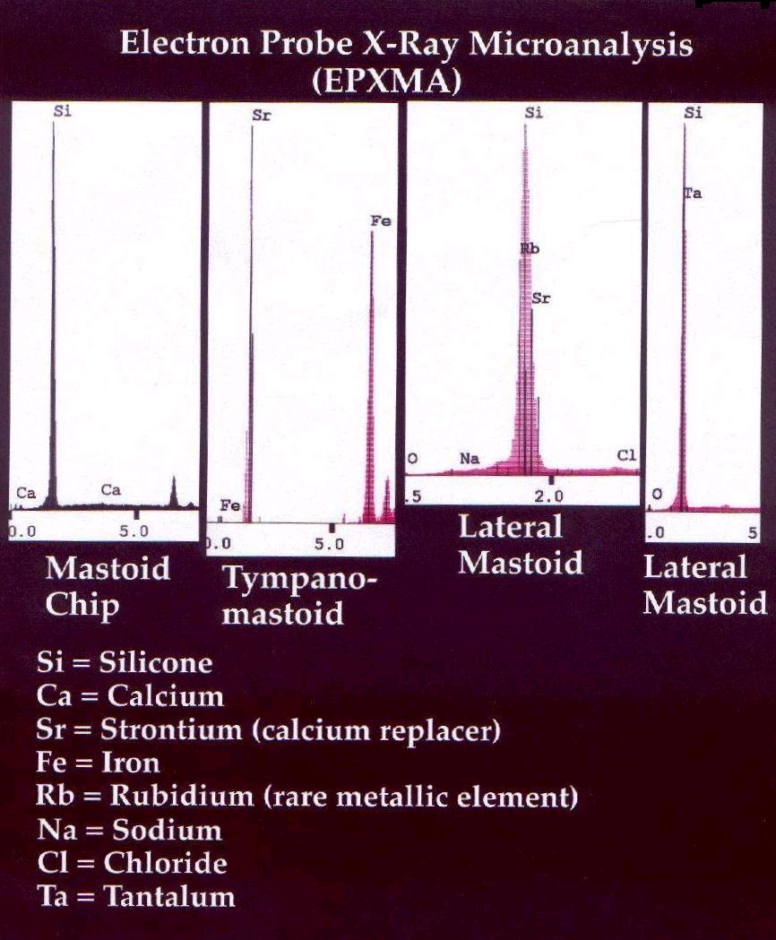
A. Electron probe x-ray microanalysis on four segments of the mastoid portion of the temporal bone from Swanscombe revealed high silicone peaks and strontium peaks, lower iron, rubidium, sodium, and chloride peaks. Calcium was also detected and although the peak was diminished in size, it passed the statistical test as set forth by Goldstein et al.*
It should be noted that strontium (Sr) is an element that replaces calcium (Ca) very readily during fossilization.
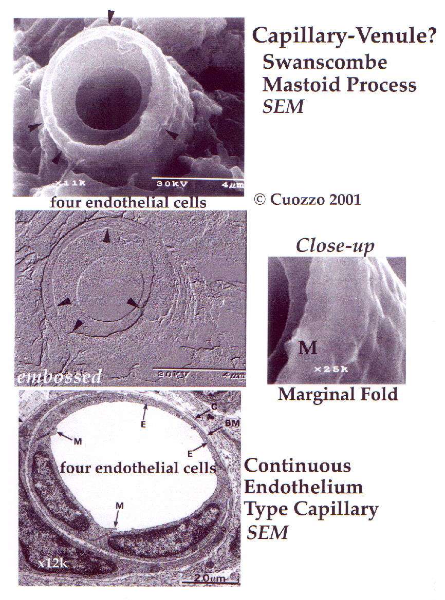
B. SEM micrograph (upper left) at 11K exposed a small blood vessel which is either a small capillary or venule in the temporal bone underlying the missing tympanic portion but anterior to the tympanomastoid fissure. The inside diameter of this blood vessel (4.4pm) is consistent with Wheater’s description *of the "smallest capillaries (3-4µm in diameter)."
Below the 11K micrograph is the same one embossed to display the divisions (marked by arrow points) between the single layer of endothelial cells that compose the wall of the vessel.
Below the embossed version of the vessel is a continuous endothelial capillary. **Note the marginal folds (M) in the 12 K slice which disclose the junctions of two cells.
The close-up seen at the right is a 25K micrograph of one of the marginal folds of the Swanscombe vessel.
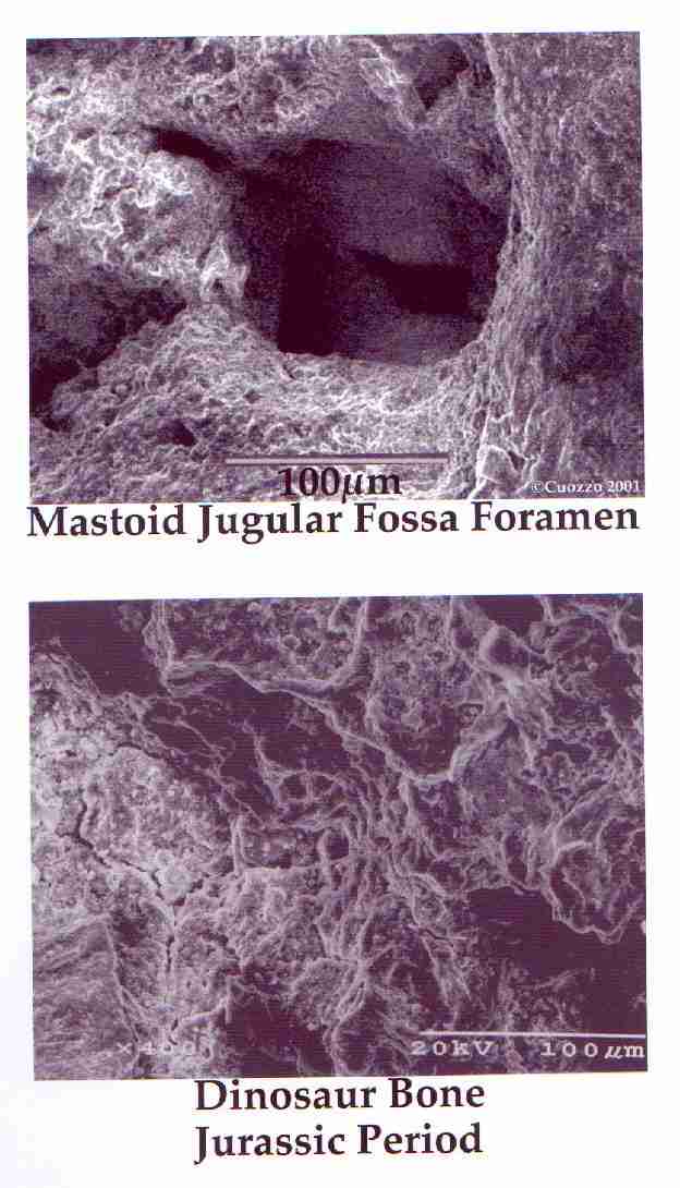
C. SEM micrographs, Upper: Foramen in the jugular fossa of the temporal bone fragment. Note the trabecular bone structure exposed on the lower edge of the foramen; compact bone on the surface.
Lower: Dinosaur bone from the Jurassic period that has a high silicone content similar to the Swanscombe temporal fragment. Note the similarity of appearance of the compact bone at the surface of both the upper and lower specimens.
D. Trichinellosis/Trichinosis
Numerous fossilized Larvae of Trichinella spiralis were found on the interfaces of the two sections of the mastoid process that were returned to me from the British Museum. The Trichenella larva diagram in the upper right displays a segmented spiral body. Similar complete segmented larval specimens on the right (1&2) 80X are just two of many complete and partial specimens of the fossilized larvae found in the Swanscombe mastoid. The lower encysted larvae are from muscle secfions*. Trichinella spiralis is found in muscles after 10 days to 4-6 weeks following ingestion of an encysted piece of animal meat. Encystment is completed in 4-6 weeks. The cycle starts with rats and rodents and continues through pigs and many species of omnivores and carnivores. The upper left two larvae are from an Alaskan Bear. **
These larvae (1&2) found in the Swanscombe woman are in the mastoid portion of the temporal bone. This means that the infestation with worms was already of long duration and that her striated muscles were also infected. In the life cycle of Trichinella, after penetration of the duodenum wall, the female worm deposits her eggs and from there the larvae spread throughout the circulatory system by means of lymphatics and capillaries to the lungs, heart and skeletal muscles. "The serous cavities and central nervous system are additional favored sites of localization although no tissue is immune.~'*** The larvae found in the mastoid, which is a well-aerated cranial membranous bony structure is evidence of extensive penetration into the Swanscombe woman’s cranium. Purulent discharge was previously reported on the posterior surface of the mastoid process.
* Encysted muscle larvae sections from Faculty of Medicine Chiang Mai University, Thailand and University of Texas, Southwestern Medical Center.
** Image Library! Center for Disease Control! Gov.
~ Robbins, S. Textbook of Pathology, W.B. Saunders Co. Phila. 1957,
373.
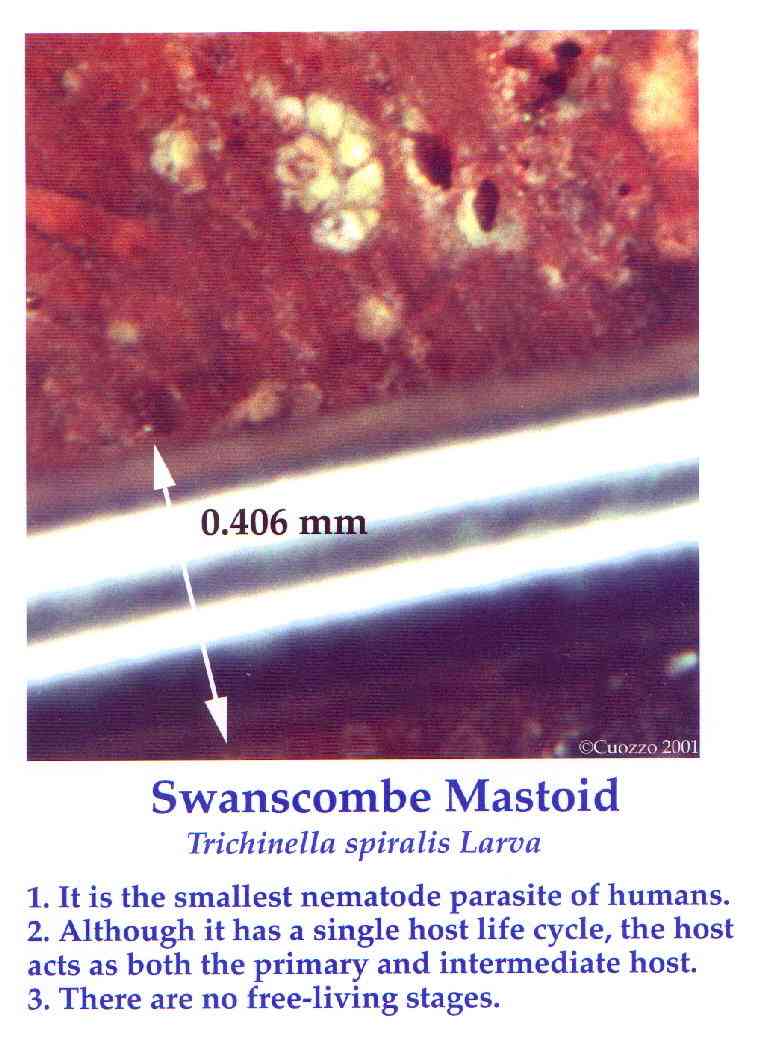
E. A third Trichinella spiralis larva found on the surface of the opposite section (lower) cut by the scientists at the British Museum. In this illustration an orthodontic wire of 0.406 mm is placed adjacent to the fossilized specimen for size comparison. Below the illustration are three characteristics of Trichinella spiralis. The most important in our case is number three. There are no free-living stages of this larva or worm. They must have a living host. They don’t live in soil or get fossilized in rocks.
Conclusions: It is my most sincere hope and prayer that we can finally put away all the discussion of whether the Swanscombe mastoid and pubis are bones. The evidence couldn’t be more definitive. There is some calcium present, the tiny blood vessel is clear, trabeculae of spongy bone are present in the jugular fossa, and there are fossilized Trichinella spiralis larvae throughout the mastoid process. These data prove conclusively that this is biologically a bone and not "worthless gravel" as determined by Dr. Chris Stringer and associates.
Due to time constraints and cost, there have been no analyses like these on the pubic bone, however in light of the overwhelming evidence presented above for the mastoid, I think it proper to include the pubis until further tests prove otherwise. But, I don’t suspect that they will, and that we must also conclude that we are dealing with heavily siliceous bones similar to those of the dinosaurs.
I wish to thank Steven Koepp Ph.D.and his graduate student Mike Ganger for the Electron Microscopic studies.
Message from Chris Stringer 1/02/02
"I got involved in the saga of Dr Cuozzo and Swanscombe because he claimed to have found more of the skeleton of the Pleistocene Swanscombe human, which is curated here: to quote from his website:
However, as the preservation of his material is demonstrably completely different from those of Pleistocene fossils from Swanscombe, including the human cranial bones, I see no reason to alter my view that he has not found more parts of the Swanscombe human.
Moreover, a colleague here informs me that the fossil within the "mastoid" gravel section is a Cretaceous foraminifera commonly found in Cretaceous flint, not (as claimed by Dr Cuozzo) a larva of Trichinella spiralis. It is, of course, quite impossible for a human bone, however fossilised, to incorporate a Cretaceous foram.
I have also invited Dr Cuozzo to come to London and make his own comparisons. However, he is apparently now arguing that his "finds" are actually something else, in which case I am more than happy to leave him to continue his own research into the constitution of the Swanscombe gravels.
Chris Stringer, Dept. of Palaeontology, The Natural History Museum, London
Dear Chris,
I am writing this to inform you that I have not changed my position on my Swanscombe discoveries. In fact, after the electron microscopy studies I am even more convinced of their validity.
I am still hoping you will understand the significance of their importance.
Sincerely,
Fri, 18 Jan 2002
"On another subject, please post a picture with an appropriate reference, that can be verified, of the fossil foram in flint from the Cretaceous Period that your collegue suggests looks like the ones in the Swanscombe
fossil. As you know, we must not judge on hearsay alone in science, and everything we do must be backed up with solid proof.
I just wish you would respect some of my findings, so we could have a decent an honorable discourse without rancor.
In parting, I wish to quote your Museum's own Theya Molleson in her chapter, "The Accumulation of Trace Metals in Bone During Fossilization" in the Book, "Trace Metals and Flouride in Bones and Teeth."
"Black, when excavating at Choukoutein , noted that many of the fragments taken from the Big Bone Bed were extensively mineralized, while others from the same stratum showed an almost complete absence of this condition. He concluded that the extent to which a fragment is mineralized or fossilized does not appear to bear any necessarily direct relation to its age."
This may very well be the case at Swanscombe.
Best Wishes for a Happy and Prosperous New Year,
Dr. Cuozzo
Click Here for an introduction to the new remains found at Swanscombe by Dr. Jack Cuozzo Swanscombe page 1
Click Here for Chris Stringers response to the find Swanscombe page 2
Click Here to Read Dr. Cuozzo's response to Dr. Stringer Swanscombe page 3
Click Here for comments from Chris Stringer, and his scans of the bones Swanscombe page 4
You are reading Swanscombe page 5
I will be posting an audio file from Dr. Cuozzo's interview with Charles Thurston regarding the new Swanscombe remains
*Goldstein, J. I., Newbury, D. B., Echlin, P., Joy, D., Fiori, C., Lifshin, B. Scanning Electron Microscopy and X-ray Microanalysis, 1st Ed. Plenum, NY, NY, 1984, 286.
*Young, B., Heath, J.W. Wheater’s Functional Histology, 4th Ed. Harcourt Publishers, 2000, 151.
* * After Wheater’ s continuous endothelial capillary.
"Dr. Jack Cuozzo, author of the book "Buried Alive - The Startling Truth about Neanderthal Man" discovered 2 additional pieces of the Swanscombe woman in September 2000".
Dr. Jack Cuozzo
1/07/02.
pg. 354-355 (Eds. Nicholas D. Priest, Frank L. Van De Vyver, CRC Press, 1990.