 |
|
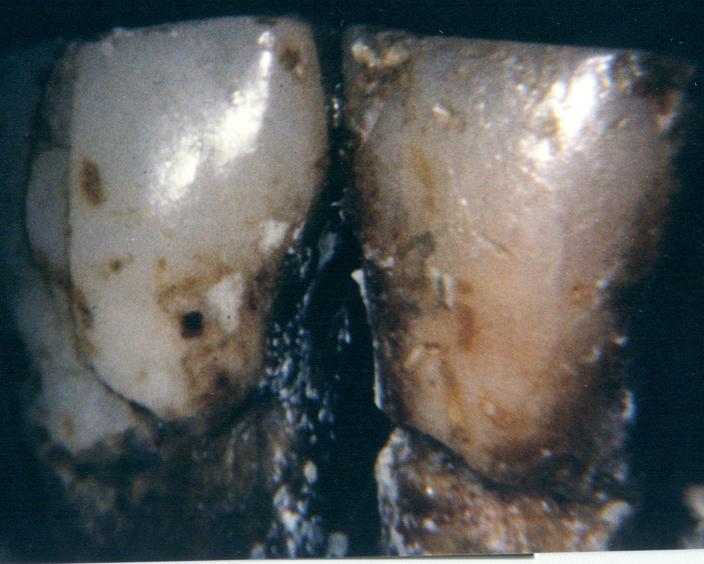
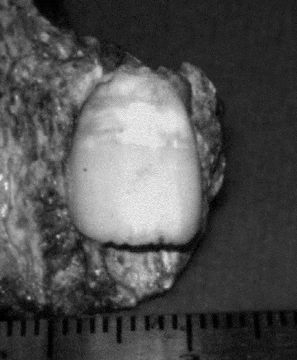
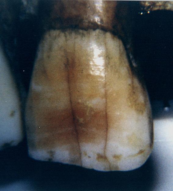
Figure 1
The lower left lateral and canine of adult Skhul V from Mt Carmel, Israel. Photographed at the Peabody Museum, Harvard University. These labial surfaces are heavily worn and demonstrate the polished nature of these teeth making it very difficult to count perikymata. In fact none are seen in the light reflection. This skull is now considered to be within H. sapiens, but very old. |
3.This is a statement I would like to challenge. (see Figure 1) I don’t think for a moment that on an incisor that is worn down to half or a quarter of its original height that the buccal or labial surface is in such a pristine condition, so that the perikymata count is EASILY done. This is a complete exaggeration. If we examine an statement by Stringer and Gamble in their book In Search of Neanderthals, (2) the so-called easy anterior tooth measurement process can be put into prospective: They state:
"The Neanderthals must have had special uses for their front teeth, for these are very large and often heavily worn compared with those of their probable ancestors. The large size of their front teeth is particularly notable considering that the rest of their dentition was relatively reduced in size (although still larger than the modern average). It is thought these teeth may have been used as a vice to hold objects other than just food items. Evidence to support this idea has come from studies of the peculiarly heavy and rounded wear patterns and damage to the front teeth: they bear microscopic traces of material, presumably both animal and vegetable, pulled outwards across the clenched teeth. Such rounded wear has even been observed on a Neanderthal milk incisor from the French cave of La Quina, showing that such activities began in early life."
Stringer and Gamble go on to describe that the front tooth damage is not isolated to France. "In an early sample (Atapuerca) and several later Neanderthals from Europe, Iraq and Israel unidirectional scratches have been observe, which suggest that something held in the teeth was being cut with stone tools."(3) Stone cutting tools can be pretty harsh on labial surfaces of teeth. It is noteworthy here to mention that 114 of the anterior teeth the authors used in this study originated from Atapuerca, without saying a word about these unidirectional scratches described by Stringer and Gamble.
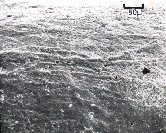
4.Continuing along these same lines of tooth wear, please observe (Figure 2) that can also be seen on (page 82) of Buried Alive. When this tooth was selected it was the most unworn permanent upper central incisor that we could find in the Harvard Peabody Collection from Mt Carmel. It is from the Tabun level B excavations. We followed the Dean and Broomage protocol carefully and scanned the labial surface of the tooth replica by means of scanning electron microscopy (SEM). It displays a rather heterogeneous surface that made it quite difficult to discern between adjacent perikymata.
 |
|
Figure 2
Scanned (SEM) labial Surface of the most unworn incisor tooth surface in the Mt Carmel collection of Harvard Peabody Museum. I made a cast of this upper central incisor using accurate silicone-based dental impression material and following the procedure recommended by Dean and Bromage in a personal communication to Dr. S. Koepp. The random elevated linear structures are the perikymata. |
 |
|
Figure 3
La Ferrassie I adult Neanderthal from France, anterior dentition. I took this photo in 1979 in the MusČe de l’Homme, Paris. Notice the extremely worn teeth (1,2.3, upper & lower) with reverse wear on the upper incisors perhaps indicating a habitual pattern of clenching a tough fibrous substance between the front teeth and pulling it upward. The same type of task may have been accomplished with the lower incisors except with a downward force applied, since they have a reverse wear pattern as well. This is called occlusal attrition ordinarily, but with normal occlusal attrition the teeth grind against each other a certain percentage of the time and so have a closer occlusion (contact) or bite (better fit). These anterior teeth have almost no fit and expose the practice of a destructive habit with a foreign object.. |
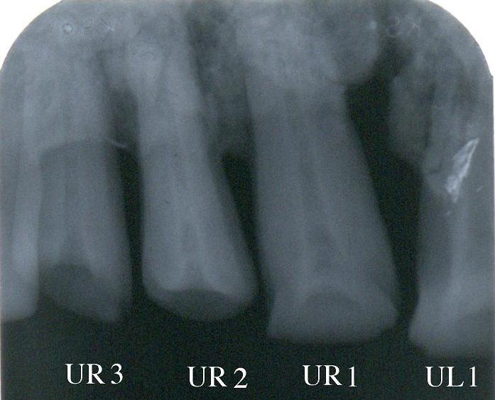
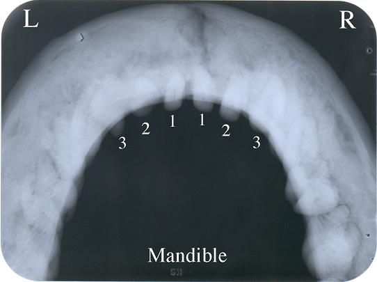
5. I include with this critique my photograph (Figure 3) of the permanent anterior teeth of one of the Neanderthals listed by Ramirez Rozzi and Bermudez de Castro: La Ferrassie. It is registered in their list of Neanderthals as contributing four anterior teeth in this study. The anterior teeth are numbered from one to three, right and left, upper and lower and there are six of them as you can see. I’d like to know which of these were the four used by the authors to count perikymata on the enamel, since there is almost no enamel left on these crowns. The lower six have absolutely none. There are tiny remnants on upper right one and two but nothing should be inferred from this miniscule amount. In any event this is only two. Where are the other two? Check the periapical radiograph (x-ray) (Figure.4) of the upper left three anterior teeth. There is not a trace of enamel on them. Also look at the occlusal radiograph (x-ray)(Figure 5) of the lower arch and there is also no evidence of any enamel on them.
 |
|
Figure 4
Periapical radiograph (x-ray) Upper left central incisor, lateral incisor and canine, La Ferrassie I. |
|
Figure 5
Occlusal radiograph of the entire lower arch of La Ferrassie I. The anterior teeth show a complete absence of enamel. |
 |
6. Would it be too much to ask of the authors to publish some of the photomicrographs that they used to calculate these numbers of perikymata? Until they do this, much of this material will remain shrouded in mystery and this is all too often one of the qualities of evolutionary science that has created an atmosphere of distrust.
7. However, let’s give them the benefit of the doubt for now and go on. Let’s assume that what they said about seeing perikymata on certain teeth where previously they had not been observed is also true. I quote from the fine print: "Preservative coating can also prevent the observation of perikymata. Teeth were carefully cleaned under microscopic inspection before direct study or moulding. This has enabled us to observe clearly perikymata even in teeth where perikymata have been reported unobserved in a previous study." What was that previous study that was unable to see what they saw? There is no footnote for this claim. Now, since no one knows who they are refuting it becomes impossible to go back and check on this other work to determine if they were correct or not. So, we have another scientific mystery.
8. Another mystery is encountered when tooth surface perikymata are counted and divided into ten sections with each section (decile) having it’s own range of perikymata.. However, we are told that the number of teeth in their Figure 1 which includes in the upper arch 52, 50, and 51 teeth respectively for Hs, Hh and Hn is only valid for the 7th to 9th deciles. This means that these 3 sections were the only ones usable in all these teeth. In the remainder of the deciles, we were not informed how many were intact in the remaining teeth. Taking just the first 52 teeth of Hs, Hh, Hn, with just three deciles totally usable, they are implying that the first 6 deciles, or 60% of the tooth crown, of these 52 teeth, is either unusable or missing. Which is it? And how many?
 |
|
Figure 6
Gibraltar 2 child upper right central incisor. Width of this tooth is 9.4 mm as opposed to 8.9 mm modern(S.D. .59 mm) male mean and 8. 67 mm (S.D. .57 mm) modern female mean. |
|
Figure 7
Gibraltar 2 child upper right central incisor crown divided in deciles or percentiles. |
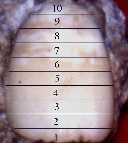 |
9. Figure 6 in this critique is the labial or buccal surface of the unerupted central incisor of the Gibraltar 2 child. (Also one of the teeth in their study) According to the authors, decile 7 has less than 15 perikymata and more than 10 by their Figure 1a chart of 26 Neanderthal central incisors or I 1. Looking at the actual decile 7 and 8 in this tooth (Figure 7) one sees very large areas of enamel hypocalcifications or opacities. This could have occurred post-mortem (after death) by an acidic decalcification in the sediments from which it was recovered, or it could have happened during the last years of this child’s life from some developmental disturbance, since the child died before the root had a chance to form. In either case the condition of this enamel surface makes difficult the possibility of an accurate count of perikymata. if it extends deeply. If it is only superficial, less than 29 microns, they still can be counted. Which is it?
10.Therefore, if we are to believe this statement, "Perikymata number in modern humans is significantly different to that in Neanderthals in all teeth from the sixth decile, except in I 1 and I 2 , where significant differences are observed from the 7th decile. Lower values are always found in Neanderthals..." Does this tooth, an upper central incisor, I 1 , fall in the category of "lower values are always found." If this is the case perhaps one of the reasons for this deficiency is the alteration in the enamel as seen in the 7th and 8th decile in Figure D.? It is a fact that light etching with dilute phosphoric acid enhances perikymata counting, but that is just light etching as I used to do in orthodontics to bond a bracket on a tooth.
11. It is necessary to challenge another statement in this report concerning the regularity of mean numbers of perikymata. in anterior teeth of Homo sapiens vs Homo neanderthalensis :..They state: "Low perikymata counts indicates that mean crown formation times were shorter in H. Heidelbergensis and H. antecessor than in Upper Paleolithic-Mesolithic H. sapiens, but Neanderthal anterior teeth are characterized by the shortest crown formation times of all these groups." Now I will quote from Buried Alive, page 82. "One more opposing viewpoint should be aired here. This was written to Nature magazine in November of 1990 by Alan Mann and his colleagues at the University of Pennsylvania,(4) Department of Anthropology, University Museum. Mann and his colleagues did another study on 12 incisors of modern man from 3000 B.C. to A.D. 800 and came up with different numbers. They found that the perikymata counts ranged from 75 to 157 (s.d. of 1-12 for individual teeth). The mean was 116. This represents a difference of 82 perikymata in their investigation." The best calculations I can make from the Romirez-Rozzi& Bermudez de Castro chart of the upper central incisor is that in deciles 6 through 9, the crucial ones, the emphasis is on the H. sapiens deciles that are packed more closely together, they say. There are approximately 79 perikymata as compared to the approximately 56 total for those deciles in H. neanderthalensis. This represents a difference of approximately 23 perikymata. (Since they give us no numbers, only charts, this is my best estimate, though I feel not far off their real numbers). One through five, they admit are fairly equal and decile number ten, we both agree is more difficult to get an accurate count due to the problems of cervical margin breakage of enamel at the edge of the tooth crown where it meets the root. (The CEJ- cemento-enamel junction) We can even allow them 10 more perkymata in decile 10 making a difference of approximately 33. Now compare this to the Mann study that found differences of 82 perikymata in 12 incisors of only one species, modern H. sapiens. If there is this much variability in our species, how can the authors assign a different species to Neanderthals that is solely based on perikymata numbers of around 33? Give them 20 more for 53. How does this compare to the Mann number of 82?
12. Then there is the huge question concerning assumptions. The first assumption is stated clearly by the authors in the text and under their Figure 1, "A 9-day interval between adjacent perikymata was assumed." This was 2 days longer than Dean et al gave Gibraltar II child in their 1986 article in the Amer. J. of Physical Anthropology, but still within modern developmental parameters. If we assume that ancient children matured at the same rates (7-10 day periods for perikymata) as modern children or even faster because of an ape heritage (Apes mature almost twice as fast as humans) then we have a uniformitarian assumption in the equation which is suspect. However, if ancient children began the decline called the secular trend towards earlier maturation that is recognized in modern science and Biblically, then they are wrong. Chapter 29 in Buried Alive details the modern secular trend toward earlier puberty and connects it to ancient historical records.
13. The purpose of an article in 2001 by Schwartz and Dean seems to clash with one of the principle statements by Ramirez-Rozzi and Bermudez de Castro. Schwartz and Dean state:"The principal aim of this study was to identify (using well-established histological methods) the developmental mechanisms that determine how male canine teeth in living great apes come to grow bigger than the canine teeth of females of the same species." They conclude: "Unlike the case for body size dimorphism, canine dimorphism in all extant large-bodied apes is the result of bimaturism, i.e., male hominoids consistently take longer to form canine crowns than do females of the same species. On the contrary, modern humans exhibit no differences in either rate or duration of canine crown, thus explaining the limited size dimorphism in this one tooth."(6) There fore if male hominoids (gorillas, both kinds of chimps, orangutans, and gibbons) take longer to develop larger canines than female hominoids it follows logically that the size of canines is time-dependent. "Significant differences in crown formation times between sexes are apparent in all ape species, and noticeably absent in modern humans."
14.From the Surprisingly Rapid Growth in Neanderthals article the statement is made: "Different numbers of perikymata do not result from differences in crown heights (Fig. 2), but indicate different crown formation times." Taking the Schwartz and Dean conclusion and this statement we can create a syllogism: a=b and b=c therefore a=c.
15. a. = differences in perikymata counts
b = differences in crown formation times
c = larger crowns
If (a) equals differences in perikymata counts (b) equals differences in crown formation times and (c)equals differences in crown formation times.
Therefore: (a) Differences in perikymata counts equals (b) differences in crown formation times,(from RR&BD) and (b) differences in crown formation times equals (c) larger crowns,(From S&D) then (a) differences in perikymata counts equals (c) larger crowns. Or larger crown heights equal larger perikymata counts. This falsifies the authors statement asserting that different numbers of perikymata do not result from different crown heights. They can result from different crown heights but they do not always by necessity have to result from different crown heights, So, they may also, result from different "packing" processes, within the various deciles. Technically, they’re off base by stating that (a) cannot equal (c). It can and does but what they also should have said is that "packing tighter deciles" can also produce more perikymata.
16.But look at this, there was reason why they made this incorrect statement. Since Neanderthals in general have larger crown heights and widths that H. sapiens, it should follow that they have more perikymata and more enamel surface than H. Sapiens.
17.From my cross- sectional crown (width x length) occlusal (biting surface) measurements of Neanderthal molars pages 312-314 in Buried Alive.
All crown measurements in square millimeters
Gibraltar 2 ……...left mand, 1stperm. molar : 152.9 (unworn)
Pech de l’AzČ …. right mand. 1st perm. molar: 156.68 (unworn)
Le Mouster 1……left mand. 1st perm. molar: 162.81 (slightly worn)
Pech de l’AzČ ….right &left maxillary perm, molars mean: 155.25 (unworn)
Le Moustier 1…...left maxillary 1st perm. molar : 162. 81 (slightly worn)
Male mand molar: 112.13 (unworn)
Female mand molar: 103.31 (unworn)
Male maxillary molar: 115.11 (unworn)
Female maxillary molar: 106.28 (unworn)
Conclusion: Neanderthals have much larger biting surface areas of their molars, therefore at least in these teeth the crown formation times should be longer. This is all dome enamel on the biting surfaces.
18.From an article entitled "Ontogeny of canine dimorphism in extant hominoids", Schwartz and Dean (7) write: "In a previous attempt to describe the ontogenetic basis of differences between male and female chimpanzee canine teeth, Dean and Beynon (1991) relied solely on surface perikymata counts in a single male and a single female canine. Results from that study suggested that male canine teeth grow at a rate different from that of females within the same period of time. It is now clear that differences in perikymata spacing result from differences in periodicity of the long-period incremental markings, and since these diverge as they approach the surface, from differences in enamel thickness (Schwartz et al., 2001).(8)
19. From Schwartz and Dean to repeat with emphasis: "It is now clear that differences in perikymata spacing result from differences in periodicity between the long period incremental markings, and since these diverge as they approach the surface, from differences in enamel thickness." The differences in periodicity is a question that has lingered in anthropology for some time. Warshawsky (1989) (9) has claimed that to continue to accept the time dependency of cross-striations is "perpetrating as great an anthropological hoax as the Piltdown Man." (10)
20. This question about periodicity and "faster extension times" must be brought into focus. The authors claim that: "the most widely spaced perikymata of all are seen in Neanderthals (fig 1b)." Stop right here and go to Figure 1b of their article and you will be surprised because you will not be able to find one photomicrograph, one close up picture, or any real proof that the authors had one piece of hard evidence that they were willing to share with their colleagues. This is not proper in science. Making enormous claims and then not being able to back them up with proof.
 |
|
Figure 8
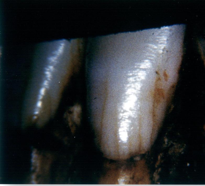
Le Mouster 1 lower left lateral incisor with light reflecting off the perikymata displaying an essentially human pattern. |
21.In contrast to this non-falsifiable practice of not showing the hard data but only charts, I would like you to examine (Figure 8) This is a relatively unworn mandibular left lateral incisor in Le Moustier 1 and it shows very clearly the perikymata packing pattern in this Neanderthal tooth. As you see the light reflecting off the perikymata lengthwise down the edge of the crown, you will notice that up high on the crown there are larger spaces between the white ridges, but as you go down further on the crown towards the root the spaces between the perikymata get smaller and smaller until they appear to merge together near the cervical margin. This is exactly what the authors say they DO NOT DO. They say they are all even spaced and not tightly packed together. Well, once again, when teeth are allowed to speak for themselves, a different story is heard.
| Figure 9
Modern human lower unerupted canine with light reflecting off the perikymata. |
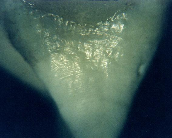 |
22. (Figure 9) is a photo with more light reflections of perikymata from the surface of the only upper central incisor of Le Moustier I. The tightly packed perikymata in the upper part of the crown also give evidence of a condition that does not exist according to the authors. This condition is very similar to that seen in (Figure 10) which is a partially formed modern human unerupted lower canine from my own collection of human teeth. This is not a cast. The light reflections are the perikymata and they become more tightly packed as they descend from the crown tip to the wider portion of this developing crown.
| Figure 10
Le Moustier 1 upper central incisor with light reflecting off the perikymata displaying an essentially human pattern.. |
 |
23.Part II to follow.
1 Perikymata: Incremental lines (Striae of Retzius) starting within enamel that are seen on the surface of the enamel as a groove between waves or ridges of the surface enamel. They have a periodicity of 8 to 10 days. The word "Peri" is taken from the Greek meaning "around" and "Kymata" is from the Greek word for "waves". This word was coined in 1895 by Preiswerk.
2 Stringer, C, Gamble C In Search of Neanderthals, Thames & Hudson Inc. NY, NY. 1993 p. 76-77.
3 Ibid
4 Mann A. Monge J. Lampl M. Dental Caution, Scientific Correspondence, Nature 348:202 Nov. 1990.
5 Schwartz G. & Dean C, Ontogeny of Canine Dimorphism in Extant Hominoids, Am. J. Physical Anthropology, 155:269-283 (2001)
6 Ibid
7 Schwartz GT, Reid DJ, Dean MC. 2001. Developmental aspects of Developmental aspects of sexual dimorphism in hominoid canines. Int J Primatol.
8 Schwartz GT Dean MC. Ontogeny of canine dimorphism in extant hominoids, Am. J. Phys. Anthro. 115:269-283, 2001.
9 Warshawsky, H. 1989. Are linear markings on dental enamel valid indicators of time? Paper presented at the annual meeting of the Canadian Association for Physical Anthropology
10 Fitzgerald CM Do enamel microstructures have regular time dependency? Conclusion from the literature and a large scale study. J. of Human Evolution (1998) 35, 371-386.